
01
More accurate diagnosis
Cone beam CT imaging provides detailed 3D views that help the team evaluate anatomy, identify concerns, and plan treatment with greater confidence.
Procedures
Cone beam CT technology gives our team precise, crystal-clear 3D images for diagnosis and treatment planning while focusing the scan on your specific area of concern.
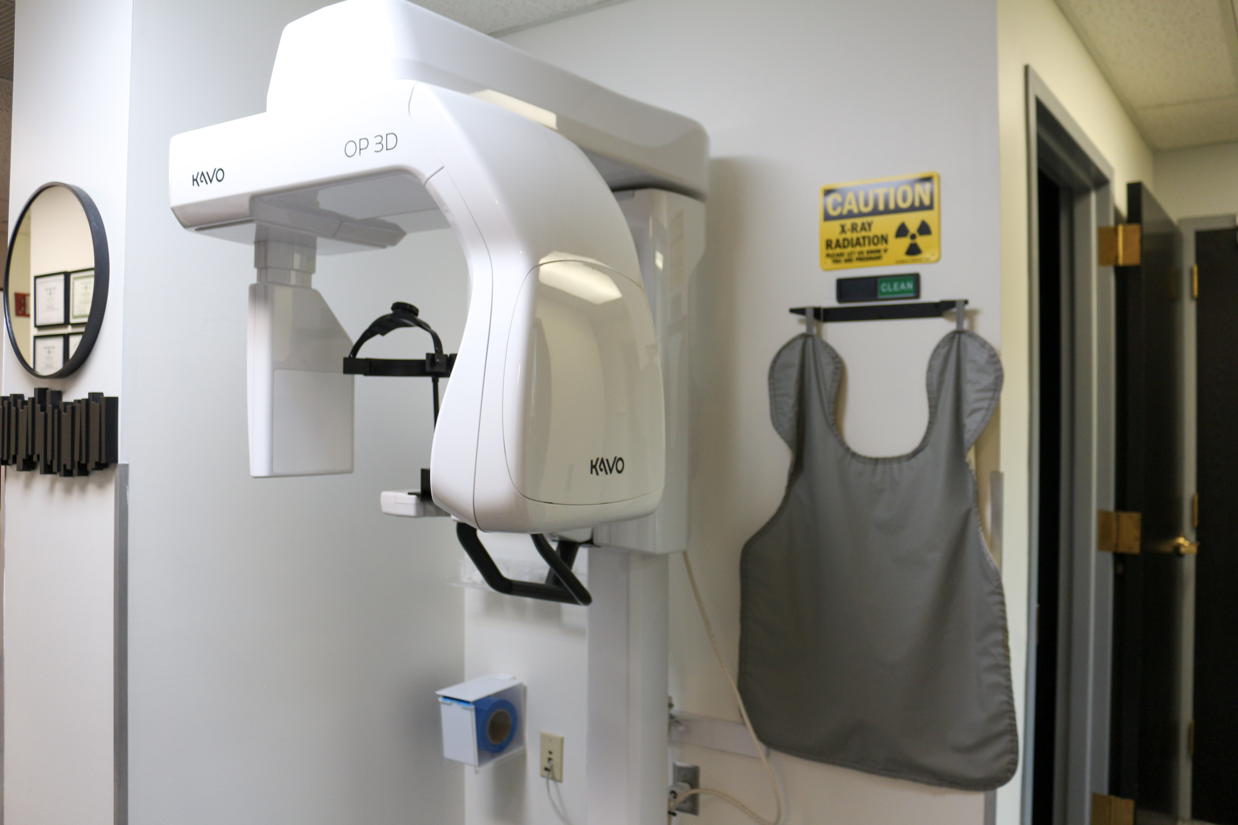
Cone beam CT scan
The CS 9300 cone beam CT system helps our oral and maxillofacial surgery team diagnose potential concerns more accurately and plan care with greater confidence.
Unlike a traditional spiral CT scanner, cone beam technology captures precise digital images while minimizing radiation exposure. The scanner can focus on the field of view that best fits your treatment needs.
The resulting 3D images help us explain your case more clearly, answer questions, and plan treatment around the actual anatomy we see.
Focused scanning
Choosing the right scanning area helps capture the information needed while limiting exposure to the area of concern.
Why 3D matters
Detailed views of teeth, bone, nerves, sinuses, and surrounding anatomy can help reduce guesswork and support clearer communication.
Cone beam CT imaging provides detailed 3D views that help the team evaluate anatomy, identify concerns, and plan treatment with greater confidence.
The scanner can focus on the specific area of concern, which helps limit radiation exposure while still capturing the needed detail.
Three-dimensional images make it easier to understand bone, teeth, nerves, sinuses, and other anatomy involved in treatment.
Images can be shared with your referring doctor so the full care team can collaborate on diagnosis and treatment planning.
Treatment planning
The scan can help evaluate anatomy before treatment and may reduce the need for multiple visits by giving the team more complete information in the office.
Dental implant planning
Wisdom tooth evaluation
Bone grafting assessment
Sinus and nerve location review
Oral pathology or surgical planning
Patient understanding
Three-dimensional visualization helps the team explain the particulars of your case and answer questions with more clarity.
Care collaboration
When appropriate, 3D images can be shared quickly with your referring dentist or doctor so the care team can coordinate treatment together.
Precise planning
We use 3D imaging to better understand your anatomy, explain your options, and plan oral surgery with confidence.